The reflex arc consists of:
– receptors that perceive irritation.
– sensitive (centripetal, afferent) nerve fiber that transmits excitation to the center
– the nerve center where excitation switches from sensory neurons to motor neurons
– motor (centrifugal, efferent) nerve fiber, carrying excitation from the central nervous system to the working organ
– effector - a working organ that carries out an effect, a reaction in response to stimulation of the receptor.
Receptors and receptive fields
Receptor- cells that perceive irritation.
Receptive field- this is the anatomical area, when irritated, this reflex is caused.
The receptive fields of the primary sensory receptors are organized most simply. For example, the tactile or nociceptive receptive field of the skin surface represents the branches of a single sensory fiber.
Receptors located in different parts of the receptive field have different sensitivity to adequate stimulation. In the center of the receptive field there is usually a highly sensitive zone, and closer to the periphery of the receptive field sensitivity decreases.
The receptive fields of secondary sensory receptors are organized in a similar way. The difference is that the branches of the afferent fiber do not end freely, but have synaptic contacts with sensitive cells - receptors. This is how gustatory, vestibular, and acoustic receptive fields are organized.
Overlap of receptive fields. The same area of the sensitive surface (for example, the skin or retina) is innervated by several sensory nerve fibers, which with their branches overlap the receptive fields of individual afferent nerves.
By overlapping receptive fields, the total sensory surface of the body increases.
Classification of reflexes.
By type of education:
Conditional (acquired) - respond to the name, the dog saliva into the light.
Unconditional (congenital) - blinking, swallowing, knee.
By location receptors:
Exteroceptive (cutaneous, visual, auditory, olfactory),
Interoceptive (from receptors of internal organs)
Proprioceptive (from receptors of muscles, tendons, joints)
By effector:
Somatic, or motor, (skeletal muscle reflexes);
Autonomic internal organs - digestive, cardiovascular, excretory, secretory, etc.
By biological origin:
Defensive, or defensive (response to tactile pain)
Digestive (irritation of oral cavity receptors.)
Sexual (hormones in the blood)
Approximate (rotation of head, body)
Motor
Posotonic (supported body postures)
By the number of synapses:
Monosynaptic, the arcs of which consist of afferent and efferent neurons (for example, knee).
Polysynaptic, the arcs of which also contain 1 or more intermediate neurons and have 2 or more synaptic switches. (somat. and veget. refs).
Disynaptic (2 synapses, 3 neurons).
By the nature of the response:
Motor\motor (muscle contractions)
Secretory (secretory gland)
Vasomotor (dilation and constriction of blood vessels)
Cardiac (measures the work of the heart muscle.)
By duration:
phasic (fast) hand withdrawal
tonic (slow) posture maintenance
According to the location of the nerve center:
Spinal (SM neurons participate) - withdrawal of the Hands from hot segments 2-4, knee reflex.
Reflexes in the brain
Bulbar (medulla oblongata) - closing of the eyelids when touching. to the cornea.
Mesencial (middle m) - vision landmark.
Diencephalic (diencephalon) – sense of smell
Cortical (BP GM cortex) – conditional. ref.
Properties of nerve centers.
1. One-sided propagation of excitation.
Excitation is transmitted from afferent to efferent neuron (reason: structure of the synapse).
Slowing down the transfer of excitation.
Conditional The presence of many synapses also depends on the strength of the stimulation (summation) and on the physical state. CNS (fatigue).
3.Summation addition of effects below threshold stimuli.
Temporary: ref. From prev. The imp has not yet passed, but the trail. Already arrived.
Spatial: mixing several. Backup They are conditioned. Images Ref.
Center of relief and occlusion.
Center relief - occurs under the action of an optimal stimulus (max response) - appears. Relief center.
When the action is min. (lower vent. Rection) occlusion has occurred.
Assimilation and transformation of the rhythm of excitation.
Transformation is a change in the frequency of a nerve impulse as it passes through the nerve center. The frequency can go up or down.
Assimilation (dance, daily routine)
Consequence
Delay in the end of a response after the cessation of stimulation. Associated with the circular nerve. Imp. By closed Circuits of neurons.
Short-term (fractions of a second)
long (seconds)
Rhythmic activity of nerve centers.
An increase or decrease in the frequency of nerve impulses associated with the properties of the synapse and the integrative duration of neurons.
8. Plasticity of nerve centers.
The ability to rebuild the functionality of a property for more effective regulation of functions, the implementation of new reflexes that were previously not characteristic of this center or the restoration of functional functions. The layer of synpses is based on a change in the malleolus structure.
Changes in excitability under the influence of chemicals.
High sensitivity to the effects of different things.
Fatigue of the nerve centers.
Associated with high synaptic fatigue. Decreased feelings. Receptors.
General principles coordination activities of the central nervous system.
Braking- special ner. percent manifested itself in a decrease or complete disappearance of the response. reactions.
The principle of convergence
Convergence is the convergence of impulses arriving along various afferent pathways in any one central neuron or nerve center.
2 . The principle of convergence is closely related to the principle common final path open Sherrinkton. Many different stimuli can excite the same motor neuron and cause the same motor response. This principle is due to the unequal number of afferent and efferent pathways.
Divergence principle
This is the contact of one neuron with many others.
Irradiation and concentration of excitation.
The spread of the excitation process to other nerve centers is called irradiation (electoral- in one direction , generalized- extensive).
The irradiation after some time is replaced by the phenomenon of concentration of excitation in the same source point of the central nervous system.
The process of irradiation plays a positive (formation of new conditioned reflexes) and negative (violation of the subtle relationships that have developed between the processes of excitation and inhibition, which leads to a disorder of motor activity) roles.
The principle of reciprocity (inhibits)
Excitation of some cells causes inhibition of others through an interneuron.
The principle of dominance
Ukhtomsky formulated the principle of dominance as a working principle of the activity of nerve centers. The term dominant denotes the dominant focus of excitation of the central nervous system, which determines the current activity of the body.
Principles of dominant focus :
Increased excitability of nerve centers;
Persistence of excitation over time;
Ability to summarize extraneous stimuli;
Inertia (the ability to maintain arousal for a long time after the end of the stimulus); the ability to cause conjugate inhibition.
The simplest reaction of the nervous system is reflex. It is a quick, automatic, stereotypical reaction to irritation, it is called an involuntary act, since it is not under the control of consciousness. Neurons that form the path of nerve impulses during a reflex act make up reflex arc. The simplest reflex arc in animals includes one neuron and has the following form:
Neuron Stimulus → Receptor - Effector → Response
This level of organization is characteristic of the nervous system of coelenterates. Reflex arcs of all groups of animals with more high level structural and functional organization consist of at least two neurons - afferent, or sensory(sensitive), conducting impulses from the receptor, and efferent, or motor(motor), transmitting impulses to the effector. Between these two neurons there may also be interneurons located in a cluster of nerve cells - a ganglion, a nerve chain, or the central nervous system (Fig. 16.13). There are a huge variety of reflexes of varying structural and functional complexity, but they can all be divided into the following four groups:
1. Monosynaptic reflexes. These are reflexes with the simplest arc found in vertebrates. The sensory neuron is in direct contact with the body of the motor neuron. Only one synapse, located in the central nervous system, participates in such an arc. Such reflexes are quite common in all vertebrates; they are involved in the regulation of muscle tone and posture (for example, the knee reflex - leg extension in knee joint). In these reflex arcs, neurons do not reach the brain, and reflex acts are carried out without its participation, since they are stereotypical and do not require thinking or conscious decision. They are economical in the number of central neurons involved and dispense with the intervention of the brain, which can “focus” on more important matters.
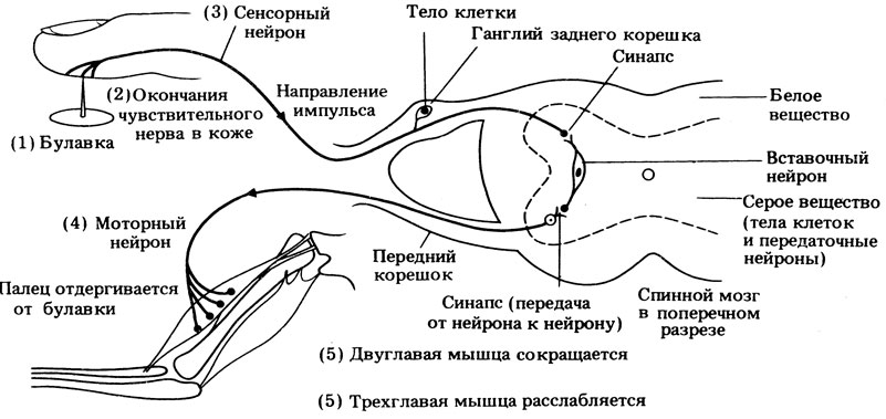
2. Polysynaptic spinal reflexes. At least two synapses located in the central nervous system are involved in such reflexes, since a third neuron is included in the arc - intercalary, or intermediate(interneuron). Synapses are present here between sensory and interneuron neurons and between intercalary and motor neurons (Fig. 16.13, B). This type of reflex act serves as an example of a simple reflex that closes in the spinal cord. In Fig. Figure 16.14 presents in a highly simplified form the reflex that occurs when a finger is pricked with a pin.
Simple reflex arcs of types 1 and 2 allow the body to carry out automatic involuntary reactions necessary to adapt to changes in the external environment (for example, the pupillary reflex or maintaining balance when moving) and to changes in the body itself (regulation of respiratory rate, blood pressure, etc.). ), and also prevent damage to the body, such as injury or burns.
3. Polysynantic reflexes involving both the spinal cord and the brain. In this type of reflex arc, a sensory neuron forms a synapse in the spinal cord with a second neuron that sends impulses to the brain. Thus, these second sensory neurons form the ascending nerve pathways (Fig. 16.15A). The brain interprets this sensory information and stores it for later use. Along with this, he is at any time this moment may initiate motor activity, and then the impulses will be transmitted by the motor neurons along the descending nerve pathway directly to the spinal motor neurons through synapses located in the same area as the output synapses of the interneurons (Fig. 16.15).
![]()
4. Conditioned reflexes. Conditioned reflexes are a type of reflex activity in which the nature of the response depends on past experience. These reflexes are coordinated by the brain. The basis of all conditioned reflexes (such as the habit of toileting, salivation at the sight and smell of food, awareness of danger) is learning (section 16.9).
There are many situations where one of two possible reflex reactions occurs, involving a particular group of muscles that can either contract or relax, which would lead to opposite results. In this situation, the usual spinal reflex would be carried out by the reflex arc shown in Fig. 16.14, however, the “conditions” under which the stimulus operates can change the response. In such cases, a more complex reflex arc operates, including both excitatory and inhibitory neurons. For example, if we grab an empty metal frying pan with our hand, which turns out to be too hot and burns our fingers, we will probably immediately let it go, but we will carefully and quickly put the equally hot food on an expensive dish that burns our fingers in its place. The difference in response indicates that we are dealing with a conditioned reflex, which involves memory and a conscious decision made by the brain. In this situation, the response is carried out along a more complex reflex pathway, shown in Fig. 16.16.
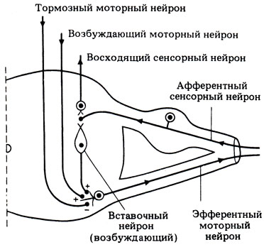
In both cases, the stimulus causes impulses that travel to the sensory part of the brain along the ascending nerve pathway. When these impulses enter the brain, it analyzes them, taking into account information coming from other senses, such as the eyes, and determines reason stimulus. The information entering the brain is compared with that which is already stored in it - with information about what will most likely happen if the spinal reflex is carried out automatically. In the case of a metal frying pan, the brain will calculate that if it is thrown, it will not cause any harm to the body or the frying pan, and will send impulses along excitatory pathway. This path goes down the spinal cord to the level where spinal cord a stimulus has arrived and forms connections with the bodies of motor neurons that carry out this reflex. The speed of impulses along this path is such that impulses from the excitatory motor neuron of the brain reach a special motor neuron simultaneously with impulses from the interneuron of a simple reflex arc. The effects of these and other impulses are summed up, and exciting impulses are sent to the muscle effector along the axon of the spinal motor neuron, causing them to throw the frying pan.
But in the case of a hot dish, the brain will quickly figure out that if you throw it, you can scald your legs, and besides, the food will be spoiled and the expensive dish will be broken. If you hold the dish and carefully place it in place, this will not cause severe burns to your fingers. After the brain makes such a decision, impulses will arise in it, which will also be transmitted to the spinal motor neurons, but this time along the inhibitory pathway. They will arrive simultaneously with excitatory impulses from the interneuron and extinguish their action. As a result, no impulses will flow through the motor neurons to the corresponding muscles and the dish will be held in the hands. At the same time, the brain can give the muscles a different program of action, and the dish will be quickly and carefully put in place.
The above description of reflex arcs is naturally greatly simplified. After all, the process of coordination, integration and regulation of functions in the body is much more complex. For example, certain neurons communicate with each other different levels spinal cord, controlling, say, the arms and legs, so that the activity of one level is coordinated with the activity of another, and another group of neurons provides general control from the brain.
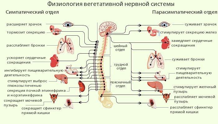
While the joint activity of the brain and the endocrine system plays an important role in the coordination of many types of nervous activity described later in this chapter, the regulation of autonomic functions is carried out by another reflex system, which is based exclusively on nervous activity. This system is called the autonomic or autonomic nervous system.
A reflex is an uncontrolled action that is the body’s response to external irritation. They can be acquired or congenital.
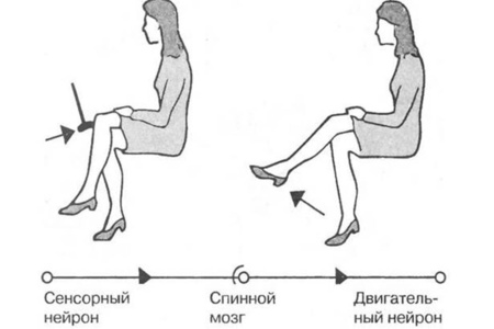
The knee reflex belongs to the group of stretch reactions or unconditioned reflexes. It can be tested by asking the patient to place one knee on top of the other and lightly but sharply hit the area under the patella (the fossa). Normally, the limb will undergo extension. The physiology of the process is based on the fact that when the muscle tendon (quadriceps femoris) is acted upon, it stretches and acts on the leg extensor muscle. This provokes spontaneous straightening of the leg. The importance of the knee reflex in ensuring the function of maintaining posture and balance.
The arc of the knee reflex
A reflex arc is the path through which a nerve impulse travels from a receptor to a muscle or organ.
The transmission scheme is based on the fact that the impulse received during an impact is transmitted from receptors (neuromuscular spindles) along axons to the bodies of sensory neurons (cells located in nodes near the spinal cord). Next, the excitation is transmitted to alpha motor neurons (in the gray matter of the spinal cord), after which the motor cells cause the quadriceps muscle to contract (the leg will “bounce”).
There is also a pathway through which the agonist muscle (flexor muscle) relaxes. The physiology is similar, the path starts from similar sensitive cells, but the signal passes through collaterals to neurons that inhibit the impulse; they are located in the lateral horns of the gray matter. From these formations the inhibitory signal goes to the motor neurons of the flexor muscle.
How to test the reflex?
The work of the neurons responsible for the occurrence of the reaction is assessed in several patient positions. You can check the reflex in the following ways:
- The patient sits on a chair, the leg being examined is crossed over the other. There is a modification when the feet are on the floor. This method described above.
- The patient is examined in a supine position, the doctor with one hand raises his leg to the level of an obtuse angle, resting on the popliteal fossa, strikes the quadriceps muscle, causing it to contract.
- The patient sits on the couch so that both legs hang from it and do not touch the floor, then the doctor acts according to the same scheme.
- The patient lies on the couch and places the knee of one leg on the knee of the other.
When carrying out this procedure, it is necessary to explain to the patient how to properly relax his legs, often inducing a knee-jerk reflex. The examination may be difficult precisely because the patient has not relaxed the muscles of the limb.
Doctors often resort to tricks by forcing the patient to solve simple problems in his head. They help distract the patient from the manipulation.
Reflex changes
There are pathological changes in the nervous system, manifested in changes in the knee reflex. Deviations may be of the following nature:
- Areflexia.
- Hyperreflexia.
A neurologist, having noted such phenomena, will conduct a comprehensive examination to make a diagnosis.
Hyperreflexia
The knee reflex is increased (hyperreflexia) - with the slightest impact on the sensitive area, the leg extends as much as possible.
Most often observed with damage to the pyramidal tracts in the anterior part of the spinal cord (anterior horns of the gray matter). The role of these structures is that they inhibit impulses that come from the brain in response to irritation.
This reflex is observed in people of the neurotic type, with some intoxications, neuritis, radiculitis, plexitis (inflammatory diseases of the nerve fibers).
The consequence of increased reflexes will be clonus - pathological movements, which are characterized by rhythmic and rapid contractions of a muscle group due to a stretched tendon. This is a chain of constant tendon reflexes. The most common manifestations are clonus of the patella and foot.

Irritation reactions with this type of pathology will decrease (hyporeflexia) or be absent (areflexia). The phenomenon manifests itself in the fact that when irritated, the knee reacts weakly or does not react at all. Pathology occurs due to a violation of the integrity and conductivity of the reflex arc at one of the stages of impulse transmission through neurons.
Most often, the absence of a reflex indicates an organic disease of the nervous system (there is a pathological formation in the brain). This may also be due to previous infection, intoxication or cachexia (loss of body weight). The listed processes lead to depletion of neurons and improper functioning of these cells. The reaction may temporarily disappear after an epileptic attack, anesthesia, or when a tourniquet is applied. With tabes dorsalis, the reflex will be absent in both legs.
In some cases, areflexia healthy people will be a variant of the norm (for diseases suffered in childhood associated with damage to the reflex arc).
Perverted reactions
A special type of knee reflex pathology is perverted or pathological reactions. They arise only when there is serious pathology nervous system.
The physiology of unconditioned reactions to the influence of a stimulus has a large clinical significance for a neurologist. The knee reflex is a mandatory element of a comprehensive examination, the condition of which indicates various diseases of the nervous system.
How to forget about joint pain?
- Joint pain limits your movements and full life...
- You are worried about discomfort, crunching and systematic pain...
- You may have tried a bunch of medications, creams and ointments...
- But judging by the fact that you are reading these lines, they did not help you much...
But orthopedist Sergei Bubnovsky claims that indeed effective remedy for joint pain exists!
Reflex and reflex arc
Reflex(from Latin “reflexus” - reflection) - the body’s reaction to changes in the external or internal environment, carried out through the central nervous system in response to irritation of receptors.
Reflexes are manifested in the occurrence or cessation of any activity of the body: in the contraction or relaxation of muscles, in the secretion or cessation of secretion of glands, in the constriction or dilation of blood vessels, etc.
Thanks to reflex activity, the body is able to quickly respond to various changes in the external environment or its internal state and adapt to these changes. In vertebrate animals, the importance of the reflex function of the central nervous system is so great that even its partial loss (during surgical removal of certain parts of the nervous system or due to diseases) often leads to profound disability and the inability to perform necessary vital functions without constant careful care.
The significance of the reflex activity of the central nervous system was fully revealed by the classical works of I.M. Sechenov and I.P. Pavlov. Back in 1862, I.M. Sechenov, in his epoch-making work “Reflexes of the Brain,” stated: “All acts of conscious and unconscious life, according to the method of origin, are reflexes.”
Types of reflexes
All reflex acts of the whole organism are divided into unconditioned and conditioned reflexes.
Unconditioned reflexes are inherited, they are inherent in every biological species; their arches are formed at the time of birth and normally remain throughout life. However, they can change under the influence of illness.
Conditioned reflexes arise with individual development and accumulation of new skills. The development of new temporary connections depends on changing environmental conditions. Conditioned reflexes are formed on the basis of unconditioned ones and with the participation of higher parts of the brain.
Unconditioned and conditioned reflexes can be classified into different groups according to a number of characteristics.
defensive
indicative
postural-tonic (reflexes of body position in space)
locomotor (reflexes of body movement in space)
exteroceptive reflex - irritation of receptors on the external surface of the body
viscero- or interoreceptive reflex - arising from irritation of receptors of internal organs and blood vessels
proprioceptive (myotatic) reflex - irritation of receptors of skeletal muscles, joints, tendons
According to biological significance
According to the location of the receptors, the irritation of which is caused by this reflex act
spinal reflexes - neurons located in the spinal cord
bulbar reflexes - carried out with the obligatory participation of neurons of the medulla oblongata
mesencephalic reflexes - carried out with the participation of midbrain neurons
diencephalic reflexes - neurons of the diencephalon are involved
cortical reflexes - carried out with the participation of neurons in the cerebral cortex
According to the location of the neurons involved in the reflex
NB!(Nota bene - pay attention!)
In reflex acts carried out with the participation of neurons located in the higher parts of the central nervous system, neurons located in the lower parts - in the intermediate, middle, medulla oblongata and spinal cord - always participate. On the other hand, with reflexes that are carried out by the spinal or medulla oblongata, midbrain or diencephalon, nerve impulses reach the higher parts of the central nervous system. Thus, this classification of reflex acts is to some extent arbitrary.
motor, or motor reflexes - muscles serve as the executive organ;
secretory reflexes - end with the secretion of glands;
vasomotor reflexes - manifested in the narrowing or expansion of blood vessels.
By the nature of the response, depending on which organs are involved in it
NB! This classification is applicable to more or less simple reflexes aimed at unifying functions within the body. With complex reflexes, in which neurons located in the higher parts of the central nervous system participate, as a rule, various executive organs are involved in the implementation of the reflex reaction, as a result of which there is a change in the relationship of the body with external environment, changes in the behavior of the body.
Examples of some relatively simple reflexes, most often studied in laboratory experiments on animals or in the clinic for diseases of the human nervous system [show] .
As noted above, such a classification of reflexes is conditional: if any reflex can be obtained with the preservation of one or another part of the central nervous system and the destruction of the overlying parts, this does not mean that this reflex is carried out in a normal body only with the participation of this part: In every reflex, all parts of the central nervous system participate to one degree or another.
Any reflex in the body is carried out using a reflex arc.
Reflex arc- this is the path along which irritation (signal) from the receptor passes to the executive organ. The structural basis of the reflex arc is formed by neural circuits consisting of receptor, intercalary and effector neurons. It is these neurons and their processes that form the path along which nerve impulses from the receptor are transmitted to the executive organ during the implementation of any reflex.
In the peripheral nervous system, reflex arcs (neural circuits) are distinguished
somatic nervous system, innervating the skeletal muscles
autonomic nervous system, innervating internal organs: heart, stomach, intestines, kidneys, liver, etc.


The reflex arc consists of five sections:
receptors, perceiving irritation and responding to it with excitement. Receptors can be the endings of long processes of centripetal nerves or microscopic bodies of various shapes from epithelial cells on which the processes of neurons end. Receptors are located in the skin, in all internal organs, clusters of receptors form sensory organs (eye, ear, etc.).
sensory (centripetal, afferent) nerve fiber, transmitting excitation to the center; a neuron that has this fiber is also called sensitive. The cell bodies of sensory neurons are located outside the central nervous system - in ganglia along the spinal cord and near the brain.
nerve center, where excitation switches from sensory neurons to motor neurons; The centers of most motor reflexes are located in the spinal cord. The brain contains centers for complex reflexes, such as protective, food, orientation, etc. In the nerve center, a synaptic connection between the sensory and motor neurons occurs.
motor (centrifugal, efferent) nerve fiber, carrying excitation from the central nervous system to the working organ; Centrifugal fiber is a long extension of a motor neuron. A motor neuron is a neuron whose process approaches the working organ and transmits a signal to it from the center.
effector- a working organ that produces an effect, a reaction in response to stimulation of the receptor. Effectors can be muscles that contract when they receive stimulation from the center, gland cells that secrete juice under the influence of nervous stimulation, or other organs.
The simplest reflex arc can be schematically represented as formed by only two neurons: receptor and effector, between which there is one synapse. This reflex arc is called bineuronal and monosynaptic. Monosynaptic reflex arcs are very rare. An example of them is the arc of the myotatic reflex.
In most cases, reflex arcs include not two, but a larger number of neurons: a receptor, one or more intercalary and an effector. Such reflex arcs are called multineuronal and polysynaptic. An example of a polysynaptic reflex arc is the reflex of withdrawing a limb in response to painful stimulation.
The reflex arc of the somatic nervous system on the way from the central nervous system to the skeletal muscle is not interrupted anywhere, unlike the reflex arc of the autonomic nervous system, which on the way from the central nervous system to the innervated organ is necessarily interrupted with the formation of a synapse - the autonomic ganglion.
Autonomic ganglia, depending on location, can be divided into three groups:
vertebral ganglia - belong to the sympathetic nervous system. They are located on both sides of the spine, forming two border trunks (they are also called sympathetic chains)
The prevertebral (prevertebral) ganglia are located at a greater distance from the spine, but at the same time they are located at some distance from the organs they innervate. The prevertebral ganglia include the ciliary ganglion, superior and middle cervical sympathetic ganglia, solar plexus, superior and inferior mesenteric nodes.
intraorgan ganglia are located in the internal organs: in the muscular walls of the heart, bronchi, middle and lower third of the esophagus, stomach, intestines, gallbladder, bladder, as well as in the glands of external and internal secretion. Parasympathetic fibers are interrupted on the cells of these ganglia.
This difference between the somatic and autonomic reflex arc is due to the anatomical structure of the nerve fibers that make up the neural chain and the speed of transmission of the nerve impulse through them.
For any reflex to occur, the integrity of all parts of the reflex arc is necessary. Violation of at least one of them leads to the disappearance of the reflex.
Reflex implementation scheme
In response to receptor stimulation, the nervous tissue enters a state of excitation, which is a nervous process that causes or enhances the activity of the organ. Excitation is based on a change in the concentration of anions and cations on both sides of the membrane of the nerve cell processes, which leads to a change in the electrical potential on the cell membrane.
In a two-neuron reflex arc (the first neuron is a dorsal ganglion cell, the second neuron is a motor neuron [motoneuron] of the anterior horn of the spinal cord), the dendrite of the dorsal ganglion cell has a significant length; it follows to the periphery as part of the sensory fibers of the nerve trunks. The dendrite ends with a special device for perceiving irritation - a receptor.
Excitation from the receptor is transmitted centripetally (centripetal) along the nerve fiber to the spinal ganglion. The axon of the spinal ganglion neuron is part of the dorsal (sensitive) root; this fiber reaches the motor neuron of the anterior horn and with the help of a synapse in which signal transmission occurs using chemical substance- mediator, establishes contact with the body of the motor neuron or with one of its dendrites. The axon of this motor neuron is part of the anterior (motor) root, through which the signal travels centrifugally (centrifugally) to the executive organ, where the corresponding motor nerve ends in a motor plaque in the muscle. As a result, muscle contraction occurs.
Excitation is carried out along nerve fibers at a speed of 0.5 to 100 m/s, in isolation and does not pass from one fiber to another, which is prevented by the membranes covering the nerve fibers.
The process of inhibition is the opposite of excitation: it stops activity, weakens or prevents its occurrence. Excitation in some centers of the nervous system is accompanied by inhibition in others: nerve impulses entering the central nervous system, can delay certain reflexes.
Both processes - excitation and inhibition - are interconnected, which ensures coordinated activity of organs and the entire organism as a whole. For example, during walking, contraction of the flexor and extensor muscles alternates: when the flexion center is excited, impulses follow to the flexor muscles, at the same time, the extension center is inhibited and does not send impulses to the extensor muscles, as a result of which the latter relax, and vice versa.
The relationship that determines the processes of excitation and inhibition, i.e. self-regulation of body functions is carried out using direct and feedback connections between the central nervous system and the executive organ. Feedback (“reverse afferentation” according to P.K. Anokhin), i.e. the connection between the executive organ and the central nervous system implies the transmission of signals from the working organ to the central nervous system about the results of its work at any given moment.
According to reverse afferentation, after the executive organ receives the efferent impulse and performs the operating effect, the executive organ signals the central nervous system to carry out the order in the periphery.
Thus, when the hand grasps an object, the eyes continuously measure the distance between the hand and the target and send their information in the form of afferent signals to the brain. In the brain there is a short circuit to efferent neurons, which transmit motor impulses to the muscles of the hand, which produce the actions necessary for it to pick up an object. The muscles simultaneously influence the receptors located in them, which continuously send sensitive signals to the brain, informing about the position of the hand at any given moment. Such two-way signaling along reflex chains continues until the distance between the hand and the object is zero, i.e. until the hand takes the object. Consequently, self-checking of the organ’s functioning is carried out all the time, possible thanks to the mechanism of “reverse afferentation”, which has the character of a vicious circle.
The existence of such a closed ring, or circular, chain of reflexes of the central nervous system ensures all the most complex corrections of processes occurring in the body under any changes in internal and external conditions (V.D. Moiseev, 1960). Without feedback mechanisms, living organisms would not be able to intelligently adapt to their environment.
Consequently, instead of the previous idea that the structure and function of the nervous system is based on an open reflex arc, the theory of information and feedback (“reverse afferentation”) gives a new idea of a closed circular chain of reflexes, of a circular system of efferent-afferent signaling. Not an open arc, but a closed circle - that’s what newest performance about the structure and function of the nervous system.
The reflex arc consists of a nerve link that perceives a signal of irritation (receptor), as well as a centripetal nerve fiber or afferent link in the form of processes of receptor neurons. These processes ensure the transmission of nerve impulses from sensory nerves along the spinal cord to the central nervous system. In addition, the reflex arc includes a central link and an efferent link, which transmits an impulse from the nerve center to the effector - an executive organ that changes its activity depending on the type of reflex.
Reflex arcs are monosynaptic, two-neuron and polysynaptic (containing three or more neurons).
Thanks to the simplest reflex arc, a person is able to automatically adapt to the slightest changes in his environment. The transmission of a nerve impulse along it allows you to pull your hand away from a hot surface in time or adjust the size of the pupil when the lighting changes. In addition, the functions of the reflex arc include the regulation of processes occurring inside the body, which makes it an indispensable condition for maintaining the stability of homeostasis (the internal environment of the body) and its constant maintenance at the desired level.
Operating principle
The resulting nerve impulse passes from the receptor along the afferent neuron to the sensitive spinal cord, where it is processed by the dendrites of the efferent neuron and transmitted to a specific gland or muscle. Most simple example The reflex reaction is the knee-jerk reflexes that occur when the therapist taps with a hammer. A polysynaptic reflex arc, which includes three or more sensory, motor and interneurons, conducts the spinal reflex not through the brain, but through the spinal cord.
The bodies of the sensory neurons of the reflex arc are located in the spinal ganglion, and the bodies of motor and interneurons are located in the gray matter of the spinal cord.
Often, the sensory neurons of the polysynaptic arc transmit information passed through the interneurons directly to the brain, which processes the received data and stores it for further use. At the same time, the nerve centers of the reflex arc can become tired, as a result of which the conduction of the impulse can weaken and even stop completely for a while - while the nerve fibers almost never get tired.