Why do we need lungs?
Breathing is a largely uncontrolled process carried out at a reflex level. A certain zone is responsible for this – the medulla oblongata. It regulates the pace and depth of breathing, focusing on the percentage of carbon dioxide concentration in the blood. The rhythm of breathing is affected by the work of the whole organism. Depending on the breathing rate, the heart rate slows down or speeds up. Physical activity causes the need for more oxygen, and our respiratory organs switch to an enhanced mode of operation.Special breathing exercises help control the pace and intensity of the breathing process. Experienced yogis can stop respiratory process for a very long period of time. This is achieved through immersion in a state of samadhi, in which vital signs are actually not recorded.
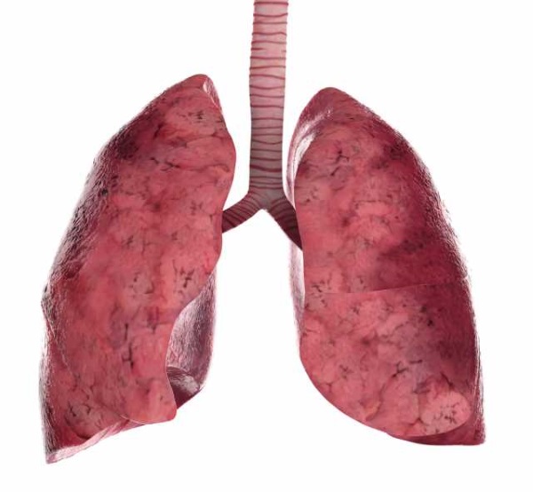
It is about 25 cm long and weighs 700 g, located in the chest cavity. The right lung is slightly larger than the left lung and is divided into three lobes; the left lung already has only two lobes. On the inside of both lungs there is an opening through which the bronchi, pulmonary arteries and pulmonary veins pass.
The lungs are more or less conical in shape and are surrounded by two membranes called the pleura. The inner pleura adheres to the pulmonary surface, while the outer pleura adheres to the chest wall. Between the pleura there is a narrow space filled with fluid. The surface tension of this fluid holds the two pleurae together but allows them to slide over one another during breathing movements.
In addition to breathing, the lungs provide an optimal level of acid-base balance in the blood, immune response, filtration of microthrombi, regulation of blood coagulation, and removal of toxins.
Structure of the lungs
The left lung has a smaller volume than the right - on average by 10%. It is longer and narrower, which is due to the peculiarities of the anatomy - placement, which is located to the left, making the width of the left lung slightly smaller.The lungs have a semi-cone shape. Their base rests on the diaphragm, and the top protrudes slightly above the collarbones.
Respiratory system Inhaled air
Can you be at school, run, eat, watch TV, sleep? it does not matter. Here you are: inhale, exhale, draw air out, send air out. Because we are made up of cells, millions of cells, and each one needs air. And when you do physical exercise activities such as dancing or playing football, your cells need more air. This is why we breathe faster and our hearts beat faster.
Respiration is the function by which living cells in the body take in oxygen and eliminate carbon dioxide. This is a gaseous exchange between atmospheric air and the body. Blood circulates within tiny vessels adjacent to every cell in the body and is the red blood cells that carry oxygen to tissues and remove carbon dioxide.
In accordance with the structure of the ribs, the surface of the lungs adjacent to them has a convex shape. The side facing the heart is concave. Thus, a space sufficient for the functioning of the heart muscle is created.
In the middle of the respiratory organ there are depressions - the main “gateways” of the oxygen transport route. They contain the main bronchus, bronchial artery, pulmonary artery, tree of nerves, lymphatic and venous vessels. The whole thing is called the “pulmonary root.”
In the lungs, red blood cells release carbon dioxide into the air and take on a new load of oxygen. The respiratory system consists of. Nasal cavities, nasopharynx, trachea, bronchial tree; which conduct, heat, humidify and filter the inhaled air of dust particles and irritating gases before they enter the lungs. The respiratory part of the lungs, formed by the lungs with respiratory bronchioles, pulmonary alveoli and elastic tissue.
The entire airway from the nostrils to the terminal bronchioles is kept moist by the presence of a layer of cells that produces a substance called mucous. Mucus humidifies the air and prevents the thin alveolar walls from drying out, as well as collecting dust particles and foreign substances. Eyelashes are types of hair on the surface of the cell that have a wave-like movement. These movements cause mucus to flow slowly into the larynx. The mucus and particles carrying the victim are then swallowed or expelled by coughing.
The surface of each lung is covered with pleura - a moist, smooth and shiny membrane. In the area of the pulmonary root, the pleura passes to the surface of the chest, forming the pleural sac.
Two deep fissures on the right lung form three lobes (upper, middle and lower). The left lung is divided by just one fissure into two parts (upper and lower lobes).
Breathing is one of the main functions of the body. It consists of supplying oxygen to the blood, oxygen that will be transferred to all cells. Without oxygen, tissues and, therefore, the entire body could not live. Oxygen is contained in the air, and the air comes into contact with the blood through a device called a "breather".
This allows the exchange between blood and air: air gives oxygen to the blood; the blood, in turn, through the lungs, leaves carbonic anhydride, which is a rejection product of cell respiration. Breathing occurs through a series of actions that allow air to pass through the airways.
In addition, this organ is divided into segments and lobules. The segments resemble pyramids, including their own artery, bronchus and nerve complex. The segment is composed of small pyramids - lobules. There can be about 800 of them per lung.
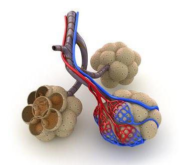
Like a tree, a bronchus penetrates each lobule. At the same time, the diameter of the “oxygen ducts” - bronchioles - gradually changes towards a decrease. The bronchioles branch and, decreasing, form alveolar tracts, to which are adjacent entire colonies and clusters of alveoli - tiny vesicles with thin walls. It is these bubbles that are the final point of transport for the delivery of oxygen to the blood. The thin walls of the alveoli consist of connective tissue, densely permeated with capillary vessels. These vessels deliver venous blood rich in carbon dioxide from the right side of the heart. The uniqueness of this system lies in the instantaneous exchange: carbon dioxide is removed into the alveoli, and oxygen is absorbed by hemoglobin contained in the blood.
Starting from the nose, this is where we get air. There are many hairs inside the nose. They serve as a filter because the air may be contaminated. And while wiping the dirt, sneeze it! Yes, this is one of the reasons why we sneeze. Expel the impurities that come with the inspired air. It will get stuck in your hair, where your body will displace a lot of air, making a storm.
The mosquito will leave at over 100 miles per hour! Air can also enter through the mouth, but in this case it is not filtered. That's why they say: a closed mouth does not penetrate a mosquito. For air, the mouth should be like a one-way street: only the exit.
With one breath, the air in the full volume of the alveolar system is not renewed. The remaining alveoli form a reserve bank of oxygen, which is used when physical activity on the body.
How do the human lungs work?
An outwardly simple “inhale-exhale” cycle in reality is a multifactorial and multi-level process.Let's look at the muscles that support the respiratory process:
From the nose or mouth, the air passes through a large tunnel full of stations, like a subway line. At the beginning of the tunnel there is a gate, a flap. This only allows air to pass through, preventing food from passing through. The first station is the larynx, which is very important for the voice. That's why we become hoarse when you have laryngitis: that's when the larynx is sick.
Next comes the vocal cords. They adjust the air when you say thick or thin. Is this the last station before reaching the lungs? or the first one when the air comes out. At the beginning, the lungs are the bronchi. We only remember these if you have bronchitis, but they are very important. The bronchi form a network through the lungs, carrying air through increasingly narrower pathways to the alveoli. Bronchitis causes these pathways to become significantly narrower, causing shortness of breath.
- Diaphragm- This is a flat muscle stretched tightly along the edge of the arch of the ribs. She separates working space lungs and heart from the abdominal cavity. This muscle is responsible for active human breathing.
- Intercostal muscles– are arranged in several layers and connect the edges of adjacent ribs. They are involved in a deep “inhalation-exhalation” cycle.

Pulmonary alveoli, the terminal station of the respiratory system. Here the air is transferred into the blood and another journey begins. For us, the main component of air is oxygen. The blood will then pick up oxygen from the red blood cells and deliver it to the outermost cells. What, this happens many times per minute.
The alveoli also contain dirty blood using air. Remember that the heart sends dirty blood to the lungs? When you breathe, cells convert oxygen into carbon dioxide. The alveoli take this used air and send it along the same path as in the bronchi, trachea, vocal cords, larynx, nose or mouth.
When you inhale, the muscles responsible for it simultaneously contract, which forces air under pressure into the airways. The diaphragm becomes flat during contraction, and the pleural cavity becomes an area of negative pressure due to the vacuum. This pressure affects the lung tissues, causing them to expand, transferring negative pressure to the respiratory and airways. As a result, air from the atmosphere enters the human lungs, since an area is formed there low blood pressure. The newly received air mixes with the remnants of the previous portion lingering in the alveoli, enriching them with oxygen and removing carbon dioxide.
Deep inhalation is achieved by weakening part of the oblique intercostal muscles, as well as contracting a group of muscles located perpendicularly. These muscles push the ribs apart, thereby increasing the volume of the chest. This creates the possibility of a 20-30 percent increase in the volume of inhaled air.
Exhalation occurs automatically - when the diaphragm relaxes. Due to their elasticity, the lungs tend to return to their original volume, squeezing out excess air. When you exhale forcefully it becomes tense muscle mass abdominals and muscles connecting the ribs.
The central element of the breathing movement is the diaphragm. This is just below the rib cage. To get air in, he lowers and pushes his stomach. To expel the air, it twitches. So when you speak, it is the diaphragm that sends air upward. Sometimes we eat and swallow air.
Then, to sob, everyone has a recipe: drink water while jumping on one leg, hold your breath and count to 83, jump with a rope while whistling the national anthem and other nonsense. The lungs are two voluminous semi-conical organs that occupy most of the chest cavity.
When you sneeze or cough, the abdominal muscles contract and intra-abdominal pressure is transmitted through the diaphragm to the lungs.
The pulmonary blood vessels emerge from the right atrium and entwine the pulmonary trunk. The blood is then distributed through the pulmonary arteries (left and right). In the lung, the vessels run parallel to the bronchi and very close to them.
Each lung has a flat base resting on the diaphragm, the muscle that separates the chest cavity from the abdominal cavity, and its upper end or apex is rounded. The inner face faces the space occupied by the center of the chest cavity, called the mediastinum, and the outer convex surface is located below the ribs.
In adults, each lung is about 25 cm high and 16 cm deep, about 10 cm wide in the right lung and about 8 cm in the left lung. The volume of the left lung is lower than that of the right because most of the heart is in the left region of the chest cavity.
The result is the enrichment of red blood cells with oxygen. Blood leaving the alveoli moves to the left side of the heart. The air entering during inhalation changes the gas composition of the alveolar voids. Oxygen levels increase and carbon dioxide levels decrease. Blood moves through the alveolar capillaries very slowly, and hemoglobin has time to attach the oxygen contained in the alveoli. At the same time, carbon dioxide is released into the alveoli.
The lungs pass through caesurae, which divide them into wolves. The right lung is counted with two caesuras that divide it into three lobes: lower, middle and upper. On the other hand, the left, slightly smaller, has one caesura and only two lobes: inferior and superior.
Each pulmonary lobe has several segments ventilated by specific bronchi: ten in the right lung and ten in the left, two of which belong to the lower lobe, forming a unit known as the uvula. Consequently, each segment is composed of numerous small secondary lobes, each containing three to five acini, small structures that correspond to the functional units of the lungs because they are where the exchange of gases between air and blood occurs.
Thus, there is a continuous exchange of gases between the atmosphere and the blood.
The main differences between the lungs of a smoker
- Healthy people have special cilia on the surface of the epithelium of the upper respiratory tract, which, with flickering movements, prevent pathogens from entering the body. Tobacco smoke damages these eyelashes, covering them with greasy soot and resins. As a result, any “infection” moves without delay into the deeper respiratory sections.
- Inflammatory processes will move further and further each time, covering all the lungs of a smoker.
- Nicotine tar (or tar) settles on the pleural surface of the lungs, which clogs the alveoli, preventing gas exchange.
- When tobacco is burned, a highly toxic carcinogen, benzopyrene, is released. It causes cancer of the lungs, larynx, oral cavity and other “smoke-conducting” organs.

On the contrary, each lung has on its inner face a large fissure, the pulmonary guild, through which the bronchi and blood vessels enter the organ. In fact, through the filaments, the corresponding main bronchi, the pulmonary arteries, which carry blood to the heart, and the pulmonary veins, which carry blood from the lungs to the heart, enter the lungs.
Once inside the lungs, the main branch of the bronchi divides into gradually smaller segments. The last branches are the terminal bronchioles, which reach the entire lung tissue. A similar process occurs with the pulmonary arteries, since from the moment they enter the lungs they are divided into thinner blood vessels until they are divided in the form of capillaries throughout the tissue of these organs.
The type of smoker's lungs depends on the person's age, length of service and place of residence. The lungs of a heavy smoker resemble black moldy cheese, chewed by worms and mice.
Tobacco smoke contains 4,000 chemical compounds: gaseous and solid particles, of which about 40 are carcinogenic: acetone, acetaldehyde, hydrogen sulfide, hydrocyanic acid, nitrobenzene, hydrogen cyanide, carbon monoxide and other extremely “useful” substances.
Frequent repeated inflammations lead to irreversible damage to the lungs. Toxins kill the “breathing tissue” of the lungs. Under the influence of resins, it is transformed into fibrous connective tissue, which is not capable of providing gas exchange. The useful area of the lungs decreases, and the volume of oxygen entering the blood is sharply reduced. Lack of oxygen leads to narrowing of the bronchi. The destructive effects of smoke provoke chronic obstruction of the lungs.
The lungs of smokers living in large industrial cities are especially affected. Their lungs are already covered with a layer of soot from automobile exhausts, emissions of combustion products and chemical reactions into the atmosphere by various enterprises.
Subsequently, the capillaries converge with each other, forming small venous vessels, which are connected in series in order to form veins with increasingly significant flow; Finally, they form the large pulmonary veins, which emerge from each pulmonary root.
Each acini, a functional unit of the lung, corresponds to a reduced amount of tissue ventilated by the terminal bronchiole. After draining into the lungs, the terminal bronchioles divide successively into thinner segments, the respiratory bronchioles, and then into the alveolar canals. At the end of each alveolar canal are the alveoli, microscopic elastic sacs with a very thin air-filled wall grouped together to form the alveolar sac.
Even if we forget about the toxic effects of tobacco smoke, one of the main symptoms - oxygen starvation - is a serious reason to think about it. Cells human body in such stressful situation age at a catastrophic rate. The heart, in a vain attempt to enrich the blood with oxygen, drains its resource many times faster. From chronic hypoxia (lack of oxygen) brain cells die en masse. Man is deteriorating intellectually.
These thin alveolar walls rest on a layer of flat lining cells surrounded by a band of supporting tissue that separates them from the adjacent alveoli, the alveolar septum. Next to the alveoli, separated only by a very thin basement membrane, are the blood capillaries that cross the lungs. There is a distance of less than 0.5 thousandths of a millimeter between the inside of one of these blood capillaries and the inside of the socket.
In the alveolar septa one can also find specialized cells in the secretion of a substance called surfactant, a liquid that coats the inner surface of the alveoli and, thanks to its physicochemical properties, prevents its collapse after expiration. This is why there are some macrophages scattered around, defense cells that detect microorganisms and other foreign particles for absorption and digestion.
Due to poor blood supply, complexion and skin condition deteriorate. The most harmless disease of a smoker may be chronic bronchitis.
Ways to improve lung health
There are widespread myths that as soon as you quit smoking, your lungs will return to their normal state within a short time. It is not true. It also takes years of normal function to remove toxins that have accumulated over the years from the lungs. Destroyed lung tissue is practically impossible to restore.Ex-smokers should follow some recommendations to get their body back to normal:
Lungs, front view and profile. They are two organs of a spongy structure and have a pyramidal shape with the base located on the diaphragm. The right is larger than the left because it has three parts or lobes, while the other has only two. Each lung consists of numerous lobes, which in turn contain the alveoli, which are the terminal extensions of the bronchi; Pleura are membranes that cover the lungs and secure them in the chest cavity.
The main function of the lung is hematosis, in which both oxygen and carbon dioxide cross the air-air barrier passively due to differences in concentration between the two phases. It is also involved in regulating body temperature. Alveoli: Tiny cavities that form the lungs on the walls of small vessels and air sacs. Outside the alveoli are networks of blood capillaries. Its walls are very thin and consist only of a layer of flat epithelial cells through which oxygen and carbon dioxide molecules easily pass through.
- Every morning you need to drink a glass of milk, as this product is an excellent adsorbent that binds and removes toxic substances from the body.
- Be proactive about taking vitamins B and C, as cigarettes were depleting your personal supply of these chemicals every day.
- Don't start doing intense sports right away. Let your body return to normal. Your worn-out heart and battered lungs will not be delighted by intense physical activity. It’s better to spend more time outdoors, walk, swim.
- Drink at least a liter of orange or lemon juice every day. This will help your body recover faster.
- Spruce shoots. It is necessary to collect young green shoots at the ends of spruce branches. It is better to collect in May or June. A layer of shoots is placed at the bottom of a liter container and sprinkled with granulated sugar. Next - again a layer of shoots and again a layer of sugar. The components fit tightly. The jar is placed in the refrigerator, after 3 weeks the shoots release juice and a sugar syrup. The syrup is filtered and stored in a cool place without access to light. Take a dessert spoon 3 times a day until the jar runs out. The drug cleanses the bronchi and lungs of toxins and “garbage”. The procedure is carried out once a year.
- Inhalation of essential oils. Boil about half a liter of water in an enamel container. Without removing the container from the flame, add a teaspoon of marjoram, eucalyptus or pine oil. Remove from heat. Next, we bend over the container and inhale the vapor for seven to ten minutes. The course period is two weeks.
- Any classes breathing exercises (especially yoga) will help your lungs cleanse and tone.
Breathe easy and be healthy!
What do our lungs look like? In the chest, 2 pleural sacs contain lung tissue. Inside the alveoli are tiny sacs of air. The apex of each lung is in the region of the supraclavicular fossa, slightly above (2-3 cm) the collarbone.
The lungs are equipped with an extensive network of blood vessels. Without a developed network of vessels, nerves and bronchi, the respiratory organ would not be able to function fully.
The lungs have lobes and segments. The interlobar fissures are filled with visceral pleura. The segments of the lungs are separated from each other by a connective tissue septum, within which vessels pass. Some segments, if damaged, can be removed during surgery without causing harm to adjacent ones. Thanks to the partitions you can see where there is a line"section" of segments.
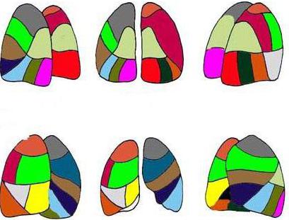
Lobes and segments of the lung. Scheme
The lungs, as you know, are a paired organ. The right lung consists of two lobes separated by grooves (lat. fissurae), and the left lung consists of three. The left lung is smaller because the heart is located to the left of center. In this area, the lung leaves part of the pericardium uncovered.
The lungs are also divided into bronchopulmonary segments (segmenta bronchopulmonalia). According to international nomenclature, both lungs are divided into 10 segments. There are 3 in the upper right lobe, 2 in the middle lobe, and 5 segments in the lower lobe. The left part is divided differently, but contains the same number of sections. The bronchopulmonary segment is a separate section of the pulmonary parenchyma, which is ventilated by 1 bronchus (namely the 3rd order bronchus) and supplied with blood from one artery.
 Each person has an individual number of such areas. The lobes and segments of the lungs develop during the period of intrauterine growth, starting from 2 months (differentiation of lobes into segments begins from 20 weeks), and some changes during development are possible. For example, in 2% of people the analogue of the right middle lobe is another lingular segment. Although most people have lingular segments of the lungs only in the left upper lobe - there are two of them.
Some people's lung segments are simply "built" differently than others, which does not mean that this is a pathological abnormality. This does not change the functioning of the lungs.
The lung segments, the diagram confirms this, look visually like irregular cones and pyramids, with their apex facing the gate of the respiratory organ. The base of the imaginary figures is located at the surface of the lungs.
Upper and middle segments of the right lung
The structural structure of the parenchyma of the left and right lungs is slightly different. The lung segments have their names in Latin and Russian (with a direct relationship to their location). Let's start with a description of the anterior section of the right lung.
- Apical (Segmentum apicale). It goes all the way to the scapular spine. Has the shape of a cone.
- Posterior (Segmentum posterius). It runs from the middle of the shoulder blade to its top edge. The segment is adjacent to the thoracic (posterolateral) wall at the level of 2-4 ribs.
- Anterior (Segmentum anterius). Located at the front. The surface (medial) of this segment is adjacent to the right atrium and the superior vena cava.
The middle share is “divided” into 2 segments:
- Lateral. Located at the level of 4 to 6 ribs. It has a pyramidal shape.
- Medial (mediale). The segment faces the chest wall anteriorly. In the middle it is adjacent to the heart, with the diaphragm running below.
A diagram of these lung segments is displayed in any modern medical encyclopedia. There may only be slightly different names. For example, the lateral segment is the outer segment, and the medial segment is often called the internal segment.
Lower 5 segments of the right lung
The right lung has 3 sections, and the very last lower section has 5 more segments. These lower segments of the lung are called:
- Apical (apicale superius).
- Medial basal, or cardiac, segment (basale mediale cardiacum).
- Anterior basal (basale anterius).
- Lateral basal (basale laterale).
- Posterior basal (basale posterius).
These segments (the last 3 basal) are largely similar in shape and morphology to the left sections. This is how the lung segments are divided on the right side. The anatomy of the left lung is somewhat different. Left side we'll look into it too.
Upper lobe and lower left lung
The left lung, some believe, should be divided into 9 parts. Due to the fact that the 7th and 8th sectors of the parenchyma of the left lung have a common bronchus, the authors of some publications insist on combining these lobes. But for now, let’s list all 10 segments:
Upper sectors:
- Apical. This segment is similar to the mirror right one.
- Rear. Sometimes apical and posterior are combined into 1.
- Front. The largest segment. It comes into contact with the left ventricle of the heart on its medial side.
- Upper lingual (Segmentum lingulare superius). Adjacent at the level of 3-5 ribs to the anterior chest wall.
- Lower lingular segment (lingulare interius). It is located directly below the upper lingular segment, and is separated below by a gap from the lower basal segments.
And the lower sectors (which are similar to the right ones) are also given in the order of their sequence:
- Apical. The topography is very similar to the same sector on the right side.
- Medial basal (cardiac). Located in front of the pulmonary ligament on the medial surface.
- Anterior basal.
- Lateral basal segment.
- Posterior basal.
Lung segments are both functional units of parenchyma and morphological ones. Therefore, for any pathology, an x-ray is prescribed. When a person is given an x-ray, an experienced radiologist immediately determines in which segment the source of the disease is located.
Blood supply
The smallest “details” of the respiratory organ are the alveoli. Alveolar sacs are vesicles covered with a thin network of capillaries through which our lungs breathe. It is in these pulmonary “atoms” that all gas exchange occurs. The lung segments contain several alveolar ducts. In total, there are 300 million alveoli in each lung. They are supplied with air by arterial capillaries. Carbon dioxide is taken up by the venous vessels.
The pulmonary arteries operate on a small scale. That is, they nourish the lung tissue and make up the pulmonary circulation. The arteries are divided into lobar and then segmental, and each feeds its own “section” of the lung. But bronchial vessels, which belong to the systemic circulation, also pass here. The pulmonary veins of the right and left lung enter the flow of the left atrium. Each segment of the lung has its own grade 3 bronchus.

On the mediastinal surface of the lung there is a “gate” hilum pulmonis - depressions through which the main veins, lymphatic vessels, bronchi and arteries pass to the lungs. This place of “intersection” of the main vessels is called the root of the lungs.
What will the x-ray show?
On an x-ray, healthy lung tissue appears as a monochromatic image. By the way, fluorography is also an x-ray, but of lower quality and the cheapest. But if cancer cannot always be seen on it, then pneumonia or tuberculosis is easy to notice. If spots of a darker shade are visible on the image, this may indicate inflammation of the lung, since the density of the tissue is increased. But lighter spots mean that the organ tissue has low density, and this also indicates problems.
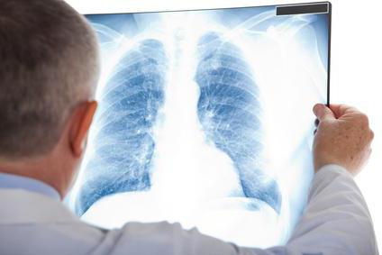
Lung segments are not visible on the x-ray. Only the overall picture is recognizable. But the radiologist must know all the segments; he must determine in which part of the pulmonary parenchyma there is an anomaly. X-rays sometimes give false positive results. Analysis of the image only provides “blurry” information. More accurate data can be obtained from computed tomography.
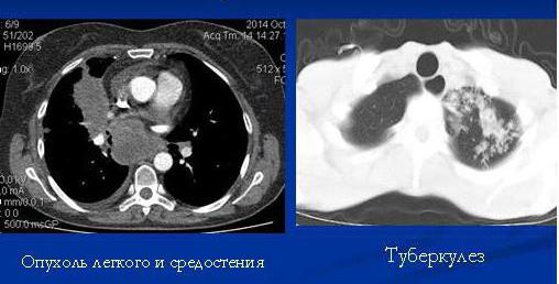
Lungs on CT
Computed tomography is the most reliable way to find out what is happening inside the pulmonary parenchyma. CT allows you to see not only lobes and segments, but also intersegmental septa, bronchi, vessels and lymph nodes. Whereas lung segments on an x-ray can only be determined topographically.
For such a study, you do not need to fast in the morning and stop taking medications. The whole procedure takes place quickly - in just 15 minutes.

Normally, a person examined using CT should not have:
- enlarged lymph nodes;
- fluid in the pleura of the lungs;
- areas of excessive density;
- no education;
- changes in the morphology of soft tissues and bones.
And also the thickness of the bronchi should correspond to the norm. Lung segments are not fully visible on CT scans. But three-dimensional picture compose and write in medical card The attending physician will view the entire series of images taken on his computer.
The patient himself will not be able to recognize the disease. All images after the study are recorded on disk or printed. And with these pictures you need to contact a pulmonologist - a doctor specializing in lung diseases.
How to keep your lungs healthy?
The greatest harm to all respiratory system caused by poor lifestyle, poor nutrition and smoking.
Even if a person lives in a stuffy city and his lungs are constantly “attacked” by construction dust, this is not the worst thing. You can clear your lungs of dust by traveling to clean forests in the summer. The worst thing is cigarette smoke. It is the toxic mixtures inhaled when smoking, tar and carbon monoxide that are scary. Therefore, you need to quit smoking without regrets.