Rotifers - the smallest multicellular creatures on Earth. Although this creature is from 0.3 to 2 mm in size, the rotifer has muscular, digestive, excretory, nervous and reproductive systems.
And the most intricate and strange method of reproduction.
“Every living thing in nature has its own characteristics and its own oddities. The most curious creatures on Earth include tiny worms, which are commonly called rotifers, and in Latin Rotifera. They are found everywhere: in large and small lakes, reservoirs, ponds, ordinary puddles and even in the smallest droplets of water on plants.And despite such prevalence, almost no one knows them: the largest rotifers barely reach two millimeters, and they are mostly microscopic in size.
Even a large rotifer is not so easy to spot in a pond. Of course, you can see it under a microscope, but to do this you need to act quickly, that is, have time to grab the rotifer with a pipette along with a drop of water, place it in the recess of a glass slide, cover it with a coverslip and try not to break it. And then you will finally see the rotifer - this extremely complex organism.
No, this is not some kind of ciliate, although the rotifer is hardly larger than it; not a single-celled creature, not a slimy lump with cilia; It looks so inconspicuous, it has approximately the same structure as a person. It has nervous system, sensory organs, muscles, glands, stomach, intestines, jaws, esophagus, kidneys, ovaries, genitals, etc. In addition, the eyes and organs of touch. And this entire complex mechanism fits in a space no larger than a comma.
But it is naturally difficult to understand everything you see without certain knowledge. K. Wesenberg-Lund in “Notes of the Academy of Sciences” (1930) describes rotifers in great detail. I will try to convey the results of his scientific research.
Rotifer cells, unlike ours, do not divide. In each organ of an animal, their number remains unchanged throughout life: cells grow, but do not multiply; damaged tissue is not restored. Asexual reproduction such as budding, as in primitive organisms, is excluded from them.
For a long time it was believed that rotifers are hermaphrodites, like snails and leeches. Scientists examined mainly females, because males were simply not noticed: they are so small that they can easily pass through the finest mesh. These reduced organisms sometimes lack important organs - for example, the digestive system. Some of the dwarf males consist almost only of a powerful reproductive system and move with the help of cilia. Their lifespan is calculated in several hours. They reproduce in a very unusual way.
The French scientist E. Maupas, in his work from 1890–1891, first noticed the presence within the same species of rotifers of three forms: one male and two female. The first of them is a microscopic “he”, extremely simplified in its structure (lives only a few hours). The second form is the eternal virgins, they lay fragile eggs and give birth to females again. And the third one lays both unfertilized eggs (also with a thin shell), from which only males develop, and fertilized ones (black, strong, adapted for wintering), which give rise to new generations of virgin females. The German scientist O. Storch called females of the first type “amictic”, and the second - “mictic” (1924).
Some rotifers have only one mating season(summer), others have two (spring and autumn). These days, tiny males dart through the water like arrows. In an aquarium, their clusters look like a whitish haze. Mating in rotifers is also unusual: the male inserts himself into the female’s body in any place he wishes. Wesemberg-Lund writes, for example, that it is quite common to see a female mating with two males, front and back. (This case was excellently illustrated by the German zoologist H. Kretschmer in the journal International Review, 1908, No. 1.)
So, first there are several generations of virgins who lay unfertilized eggs; when there are a lot of them in the reservoir, other females hatch, laying both unfertilized eggs (from which males develop), and fertilized ones - more hardy, capable of overwintering - which again supply virgin females.
Yes, you can hardly find stranger reproduction in nature.
Rotifers, of course, are for the most part simply invisible to us. However, one should not forget about these creatures when writing about life in a pond."
(c) Hans Scherfig "Pond"
Some ciliates-slippers are an order of magnitude larger than rotifers, and sometimes multicellular rotifer runs the risk of being devoured by a single-celled ciliate!
The natural existence of these giant cells in deep ocean trenches expands our knowledge of the biological diversity of living organisms on the planet.
Unlike multicellular organisms, the smallest of which can still be seen with the naked eye, most single-celled organisms are so small that they can only be seen with a microscope. However, among them there are also real giants of the microworld. For example, amoebas grow up to 0.3 millimeters, and ciliates - slippers up to 3 mm. But recent scientific discoveries have proven that such dimensions are far from the limit for the simplest organisms. Just look at the discovery of an amazing xenophyophora.
The natural existence of these giant cells in deep ocean trenches expands our knowledge of the biological diversity of living organisms on the planet and their ability to adapt to survive in extreme environments.
Xenophyophores today are perhaps one of the deepest-sea single-celled organisms. Before this, they were encountered at a depth of about 7,000 meters. But while exploring the Mariana Trench in 2011, researchers came across this microorganism at an incredible depth of 10,700 meters! The scientific world was incredibly amazed by this find!
Xenophyophores, as is currently known, can reach a diameter of 10 centimeters and serve as a habitat for a variety of multicellular animals. They were first described by biologists back in 1889, but due to mistake and insufficient information about the animal, they were classified as sponges. Fortunately, modern research showed that xenophyophores consist of cytoplasm and evenly distributed nuclei. This means that they belong to the type of simplest single-celled organisms - foraminifera. However, their appearance can be quite varied. Some are disc-shaped, others are sponge-shaped, etc.
Meanwhile, a detailed study of the life and structure of xenophyophores is very complicated, since their habitat of this animal is quite inaccessible due to the extremely unfavorable conditions environment. In addition, the extreme fragility of their body, samples of which were taken for research, is immediately destroyed and becomes useless for further study.
From the exact data known to us, we can say that xenophyophores are the largest single-celled organisms in nature today. Due to the characteristics of their habitats, the animal is highly resistant to low temperatures And high blood pressure water column at great depth. Their bodies also contain a lot of lead, uranium and mercury, which are extremely toxic to ordinary living cells. It is believed that xenophyophores feed by processing and filtering mud. Here they find various benthic microorganisms and, like amoebas, envelop their prey with pseudopods.
An ostrich egg, reaching 15 cm in height and weighing about 1.5 kg, is often cited as an example of the largest cell of living organisms, but this is a myth.
Contrary to popular belief, there are at least a few other types of living cells that are larger than an ostrich egg. It is possible that ostrich eggs may be the heaviest cells in nature, but tests have not yet been carried out.
If we talk about size and not weight, then an ostrich egg is not the largest cage. Much larger nerve cells of large animals like giant squid- their nerve cells can reach 12 meters in length, which is about 80 times larger than an ostrich egg.
Children raised by animals

10 mysteries of the world that science has finally revealed 2,500-Year-Old Scientific Mystery: Why We Yawn Miracle China: peas that can suppress appetite for several days In Brazil, a live fish more than a meter long was pulled out of a patient The elusive Afghan "vampire deer" 6 objective reasons not to be afraid of germs The world's first cat piano Incredible shot: rainbow, top view 10 Attempts to Explain the Existence of Life Without Darwin's Theory of Evolution


Despite the fact that most people eat the very obvious eggs of birds and fish almost every day, the words “single-celled organism” conjure up something that can only be seen through a microscope. Indeed, the vast majority of single-celled creatures do not exceed dimensions of hundredths of a millimeter, and this can be explained by a number of factors. It is more difficult for large living cells to maintain structural integrity, it is more difficult to transport food and waste within the body, in addition, impressive growth requires a fair amount of energy, which is evolutionarily disadvantageous.
But the world of microbes is rich in species, old and diverse, and therefore full of exceptions to the rules. And some organisms, to which the prefix “micro-” would be attached, despite the evolutionary benefit, do not achieve anything at all. Which, naturally, delights and fascinates.
Trumpeter ciliate
This freshwater creature resembles the trumpet of an ancient gramophone and grows up to 2 mm in length, so the trumpet ciliate can be studied without instruments. Protozoa of the genus Stentor are well known to microbial enthusiasts. Two millimeters does not seem super long, but many of nature's multicellular children take up much less space in their habitats and on glass slides.
What makes the trumpeter ciliate a colossus in the world of small fry is its anatomy. Unlike ordinary eukaryotes, Stentor contains not one, but several nuclei. It makes it easier for him daily work to keep yourself in spirit. In the case of this ciliate, numerous small nuclei are responsible for reproduction, and the large nucleus - the macronucleus - manages everything else, playing the role of a kind of brain center.
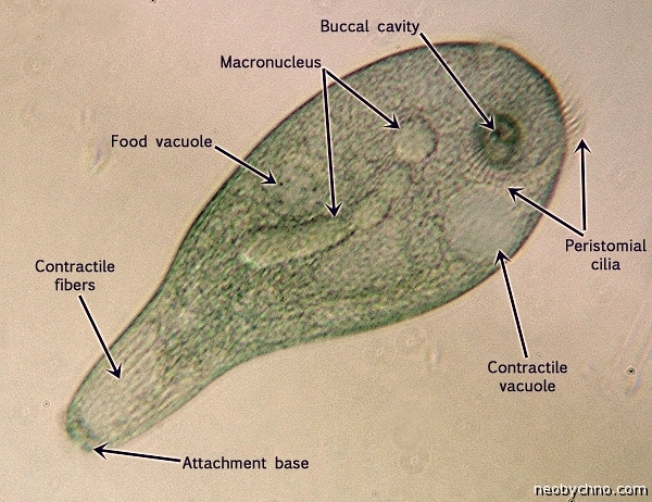
The body of the trumpeter is covered with cilia of different lengths. Their friendly movements allow the ciliates to swim. These microcosm colossi feed on, for example, silt. The function of the mouth is performed by the narrow end of the “pipe”. At the same time, some bacteria, small protozoa, and even tiny unlucky multicellular organisms end up in the food.

Bahamas Thunder
One day, scientists from the University of Texas went to the bottom of the sea near the Bahamas and discovered there, in the gloomy depths, dozens of unusual spherical objects the size of grapes. These objects seemed motionless, but clearly left traces in the sand up to half a meter long. At first, experts thought about some unknown mollusks or even strangely behaving poop. The truth was amazing, because the mysterious piles turned out to be spherical protozoa with a diameter of up to 3 centimeters. Which rolled along the bottom of the sea in almost zero temperature water.

The Bahama Gromya is an amoeba-like organism with a shell that is soft and porous. Pseudopodia are inserted into the holes in it, with the help of which the gromia moves along the bottom, feeding on organic matter caught along the way.
The discovery of this creature changed some views on the evolution of living beings, since it was previously believed that multicellular animals with bilateral symmetry were the first to learn to crawl back in Precambrian times. And the traces that gromia leaves are very similar to ancient fossilized prints that are almost 2 billion years old.
Unfortunately, little is known about these cytoplasmic balls because it is very difficult to get live Gromia specimens into the laboratory. Despite their shells, protozoa are very fragile and vulnerable. Scientists say that they are much softer than grapes, which these giant microbes are somewhat similar to.
Acetabularia
Known as the “mermaid glass,” Acetabularia is a unique genus of green algae similar in shape to cap mushrooms. These plants of shallow tropical seas are up to 10 cm in length and usually grow in groups, attaching their legs to bottom stones and showing off their light green caps.

Typically, large single-celled creatures have more than one nucleus, which is not the case with the amazing Acetabularia, which spends most of its life with just one giant DNA container located at the base of its “stalk.” Only at the hour of reproduction are additional nuclei formed, migrating to the top of the algae, where they turn into spore-like cysts, which, after wintering and complex transformation, become young acetabularia. Life cycle These colossal coenocytes are about three years old.

In experiments carried out with Nazi money in the 1930s and 40s by German scientist Joachim Hammerling, it was found that after one species of acetabularia is transplanted with the nucleus of another species of algae, the original plant begins to form a new cap, transforming into an unusual hybrid.

In addition, the “glass from which mermaids drink” perfectly regenerates when damaged, which is very reminiscent of some multicellular species of the world of flora and fauna.
Bellied Valonia
Some call this funny shallow-water creature “the eye of a sailor,” others simply call it “a bubble algae.” Valonia potbellied easily grows up to 4 cm in diameter and even more, one organism - one living cell with many nuclei, most often geographically solitary and always looking like a polished greenish pebble. Sometimes small “multicellular organisms” also take root on the surface of this unicellular marine miracle.

Despite the biological strangeness and exotic appearance of the algae, the pot-bellied wallonia is not favored by the owners of large marine aquariums. If a plant accidentally invades, it will take over the entire bottom, making it terribly difficult to get rid of. Crushing or tearing this tenacious weed into pieces is not the case, because it is through cell division that the pot-bellied wallonia with its “collection” of nuclei reproduces.


Caulerpa thyssolifolia
You might think about it as if it were some kind of fern, but in essence this plant is much simpler. And much more decisive in growth. What appears to an inexperienced diver to be thickets of underwater flora will actually turn out to be one or just a few living cells, “masquerading” as complex multicellular bushes. These primitive creatures are called "caulerpa taxifolia", or simply caulerpa herringbone, an amazing creeping thyssolid stem. One cell of this green algae, with its countless DNA stores, can very quickly expand almost three meters in width, which regularly happens in the Mediterranean Sea, destroying the healthy ecology of the depths there. For this reason, the herringbone caulerpa is recognized as a particularly harmful weed. In California, this “giant microbe” is generally considered an illegal species.

The Mediterranean variety of thyssolist caulerpa, whose cells reach record sizes, owes its status as a pest to humans. Just half a century ago, this unusual algae did not live at all in the Mediterranean Sea. But in the 1970s, an aquarium in Germany ordered specimens of caulerpa from the tropics, but not just for beauty and easy care. Inquisitive Germans subjected the “Christmas tree” to technical abuse. The macrophyte was irradiated with ultraviolet light and treated with chemical mutagens. The result was a single-celled monster, growing very quickly and resistant to low temperatures. The cold-resistant and attractive-looking algae was released into the Mediterranean Sea in 1980 - some amateur aquarist from Monaco tried.

In four years, the inevitable happened. After escaping from the aquarium, the mutated caulerpa victoriously occupied the coastal waters of the Mediterranean. Unlike its natural counterpart, the mutant cell turned out to be not only aggressive, but also resistant to pollution. Moreover, it is capable of regenerating from a piece only a centimeter in size. And poisonous. Attempts to clear the resort's shallow waters of caulerpa thickets failed.

Therefore, at the end of the 20th century, the nickname “killer algae” was assigned to the single-celled organism “Caulerpa taxifolia”. The plant is included in the hundred most dangerous invasive species, stopping the spread of which is the sacred duty of every concerned earthling.

Amoeba Chaos
Imagine an amoeba from a school textbook. Enlarge it to the size of a sesame seed. You will get the creature Chaos carolinensis. Since such protozoa constantly change shape, the champions among chaos are able to stretch up to 5 mm in length. Such heavy single-celled organisms can be fatally wounded simply by covering them with a microscope slide.

Despite its impressive size, Chaos carolinensis behaves in the same way as its microscopic pseudopod-bearing relatives. With the help of pseudopodia, chaos move, and they also grab food. The food in the vacuoles is then digested alive, and the remaining waste is thrown out of the cell to the outside. The huge amoeba feeds on microbes of other species, as well as small animals such as cladocerans. Chaos will eat almost non-stop until it is ready to reproduce.

Like its neighbors on the list of giants of the microbial world, unicellular chaos has many control centers, simply because one nucleus is not able to control such a massive cell. Depending on size, Chaos carolinensis can have up to 1000 nuclei.

Spirostomum
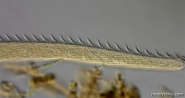
The ciliate spirostomum can be found and seen in both fresh and salt waters. And mistaken for some little worm. The elongated body of the spirostomum reaches a length of 4 millimeters. Only when looking through the microscope eyepiece does it become clear that this mobile creature is one large and very long cell, covered with a dense forest of cilia.

Spirostomum is the champion of the microbial world in its ability to change body volume. When disturbed, a ciliate can shrink by 75% in less than 1/200 of a second - faster than any other living cell.

Unlike the voracious trumpet ciliates, Spirostomum does not eat multicellular creatures, but only gets by on bacteria. Giants reproduce by simple division and really don’t like it if there are heavy metals in the water, which makes these ciliates friends of ecologists.
Siringammina is the most fragile
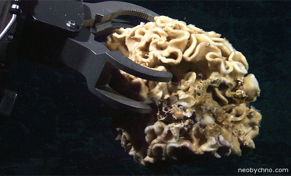
Another useful candidate for the title of the largest single-celled creature on Earth is a fragile “monster” from the xenophyophore class. This class of “carrying other people’s bodies” organisms includes many inhabitants of the ocean floor, clumps of cytoplasm that build for themselves eternal night fragile wicker “houses” made from the remains of other creatures, for example, sponges or radiolarians. Xenophyophore cells make construction glue themselves, following commands coming chemically from numerous nuclei that float in massive clumps of cytoplasm. The largest of these clumps reaches 20 cm in size, is readily colonized by worms and bears the specific name Syringammina fragilissima.

Unfortunately, the life and biology of syringammina (“sand flute of Pan” in translation) is still little studied. Scientists suspect that this single-celled bacteria feeds, but no one has seen what the process itself looks like. There is an opinion that the fragile syringammina grows microbes for its diet within itself. The mechanism of reproduction of these rhizaria is also unclear.

The fragile deep-sea creatures were discovered in 1882 by the Scots, off their native North Sea shores. Subsequently, syringammin was found on the shelf of northern Africa.
Their name is legion...
Among the terrestrial unicellular giants special attention deserved, of course, meter-long slime molds, inhabitants of dead wood. Which at first for a long time mistaken for mushrooms.

However, slime molds (in particular, multi-headed Fusarium) turned out to be not only more primitive, but also in some ways much smarter than mushrooms. You can read about the interesting conclusions of Japanese scientists in this regard in the material.
We are accustomed to thinking that single-celled organisms can only be seen under a microscope. However, almost everywhere on the bottom of the World Ocean, where there is little oxygen and where there is no sunlight at all, giant single-celled organisms, such as xenophyophores, live.
Representatives of the species Syringammina fragilissima, belonging to this class, can reach 20 centimeters in diameter, which makes them the largest single-celled animals on Earth
Xenophyophores were first described in 1889 and classified as sponges. But only recently have scientists classified them as a type of simplest single-celled organisms - foraminifera. Xenophyophores consist of cytoplasm and numerous nuclei evenly distributed in it. These organisms have a variety of appearance. For example, individuals of some species may have the shape of a disk, tetrahedron, or sea sponge. 
Xenophyophores take root on the bottom covered with silty sediments. In some places their numbers can be higher than 2000 individuals per 100 m². These giant protozoans are thought to feed like amoebas, enveloping their food in special growths called pseudopods. As with all detritivores, xenophyophores feed on dead organic matter, namely bottom sediments. 
Nowadays, xenophyophores have been studied rather poorly due to their inaccessible habitat - some species live at the bottom Mariana Trench- at a depth of more than 10,000 meters. The second factor is their extreme fragility. When scientists take samples for study, they invariably break down, making these organisms useless for study outside their habitat. 
Nevertheless, it is already known today that xenophyophores are an important part of benthic ecosystems, since they help maintain biological diversity in them. These organisms constantly recycle sediments on the bottom, thereby providing habitat for other organisms. Studies have shown that in places with a large number of xenophyophores, 3-4 times more crustaceans, echinoderms and mollusks live than in areas where these unicellular organisms are absent. 
Interestingly, in addition to xenophyophores, there are other single-celled organisms that can be seen with the naked eye:
Valonia potassium is a type of green algae. The shape can vary from spherical to oval, color from grass-green to dark green. In water it can appear silver, sea green or even blackish. The intensity of the color is determined by the number of chloroplasts in the cell. The surface of the algae is mirror-shiny, like glass. 


Acetabularia is a genus of green algae. The stem of an adult plant has a length from 2-3 cm to 4-6 cm, and the cap (umbrella) is up to 1 cm in diameter.
It has interesting ability to the regeneration of all lost parts, except the cell nucleus. Moreover, the only nucleus of this single-celled plant is located in a rhizoid (pedicle) attached to the stones. 
Caulerpa is a genus of marine green algae, which is a complex of cells devoid of intercellular septa, therefore it is a single cell with numerous nuclei, and can reach a length of 2.8 m, which allows us to consider them the largest single-celled organism in the world, with a reservation of course.